Confocal laser endomicroscopy for superficial esophageal squamous cell carcinoma
发表时间:2019-12-18 10:28
Background and study aims: Confocal laser en− domicroscopy (CLE) allows subsurface imaging of gastrointestinal mucosa in vivo. The goal of the present study was to compare the endomi− croscopic characteristics of cells and intrapapil− larycapillary loops (IPCLs) in normal and superfi− cial esophageal squamous cell carcinoma (SESC). Patients and methods: We recruited consecu− tive patients with SESC diagnosed byconvention− al endoscopy and confirmed by histopathology between July 2006 and May 2008. The confocal endoscopic images of these patients were collec− ted and compared with the corresponding histol− ogy. The characteristic patterns of cells and IPCLs was then analyzed from these images of malig− nant and normal mucosa. The quality of images and interobserver variations of two endoscopists were also evaluated. Results: Overall, 64 samples from 57 subjects (27 SESCs, 30 controls) were examined by CLE. The confocal images corresponded to the hema− toxylin and eosin staining from the same sites. The confocal images showed that there was a sig− nificantly higher proportion of squamous epithe− lial cells with irregular arrangement (79.4% vs. 10.0%, P<0.001), increased diameter of IPCLs (26.0?m vs. 19.2?m, P<0.001), and irregular shape IPCLs (82.4% vs. 36.7%, P= 0.0002) in the SESC group compared with the controls. Massive IPCLs with tortuous vessels (44.1% vs. 0%, P <0.0001), and long branching IPCLs (23.5% vs. 3.3%, P= 0.0204) were frequently observed in the SESC group. In this study, about 35.5% of ima− ges were graded as good quality, and the interob− server agreement for the prediction of cancerous mucosa was graded as substantial. Conclusions: CLE can be used to distinguish can− cerous from normal epithelium, which gives it potential value for early detection of esophageal carcinoma. The difficulty in obtaining good ima− ges in the esophagus by CLE is a latent problem.
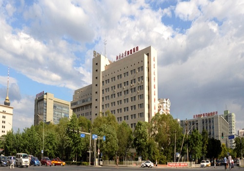
第一署名医院:山东大学齐鲁医院;北京世纪坛医院
Confocal laser endomicroscopy for superficial esophageal squamous cell carcinoma (查看pdf)